¹Department of Ophthalmology and Vision Sciences, University of Toronto
A 67-year-old man presented with acute photopsia in his right eye. Clinical examination revealed a visual acuity of 20/25, and a superior retinal break, which was treated with laser retinopexy.
Incidentally, macular retinal pigment epithelial changes were noted. A spectral domain OCT was obtained which revealed a shallow and irregular elevation of the retinal pigment epithelium (SIRE), also known as the double layer sign (see Figure 1).
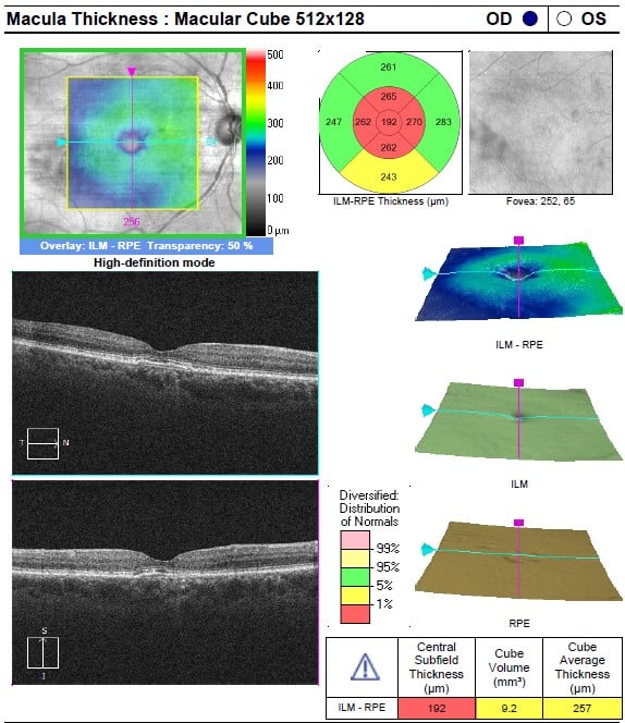
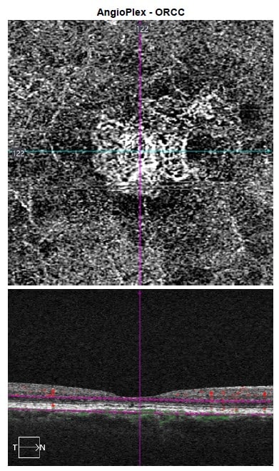
This finding, which represents a separation between Bruch’s membrane and the hyperreflective retinal pigment epithelium above it, can be suggestive of subclinical, nonexudative macular neovascularization (MNV). OCT angiography was obtained which confirmed the presence of a type 1 MNV (See Figure 2).
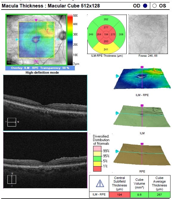
This eye was monitored with serial OCT scans. After 5 years, the visual acuity remained stable at 20/25 and no evidence of exudation or geographic atrophy was noted (see Figure 3).
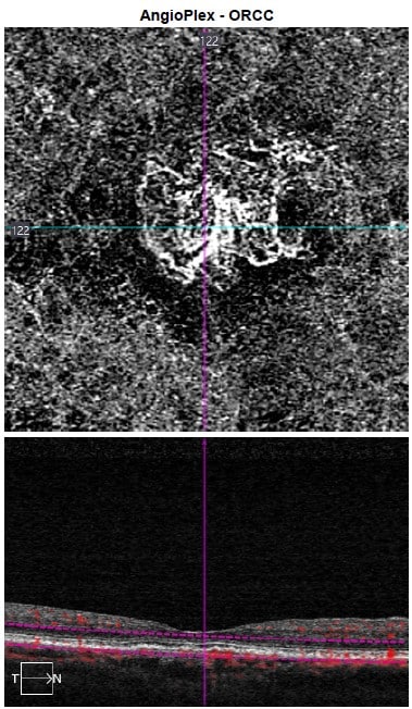
OCT angiography showed maturation of the MNV, with pruning of branching vessels and no change in the overall lesion size (see Figure 4).
SIRE was originally reported in PCV but can be find in a myriad of retinal diseases, including AMD, CSCR, and pachychoroidal disease.1 OCT angiography is very helpful in detecting subclinical MNV, with en face imaging nicely highlighting suspect areas. It has been hypothesized that type 1 MNV may support overlying RPE and photoreceptors, protecting against macular atrophy.2 This case highlights that nonexudative MNV can be stable overtime and can be safely monitored, deferring anti-VEGF therapy only if exudation occurs.