¹ Department of Ophthalmology and Visual Sciences, Dalhousie University, Halifax, NS, Canada ² Department of Ophthalmology and Visual Sciences, University of Alberta, Edmonton, AB, Canada

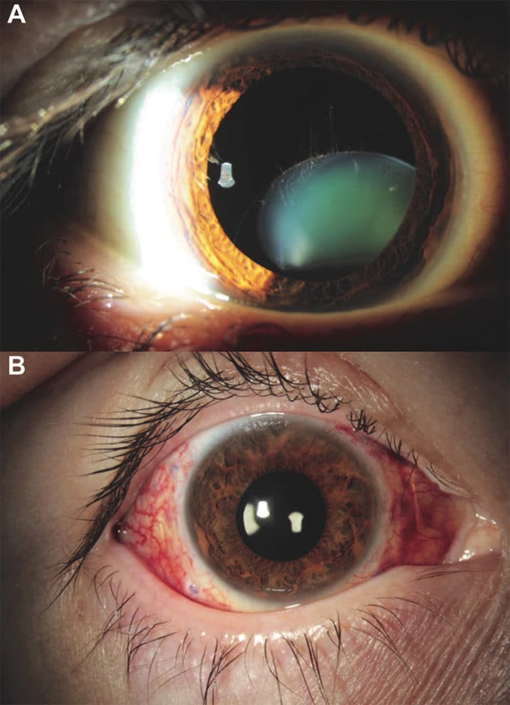
A 56-year-old male presented with decreased vision in the right eye secondary to ectopia lentis. The anterior segment photograph of the right eye captures the subluxing phakic lens. A few remaining intact, but stretched, zonules are visualized to be valiantly hanging on to the descending lens (Fig 1A).
The patient underwent a pars plana vitrectomy, pars plana lensectomy, and scleral suturing of an Akreos AO60 intraocular lens (Bausch & Lomb Bausch, Rochester, NY) with Gore-Tex suture (W.L. Gore & Associates, Newark, DE) (Fig 1B). His visual acuity improved from 20/150 pre-operatively, to 20/25 without correction post-operatively.