¹ Department of Ophthalmology and Visual Sciences, Dalhousie University, Halifax, NS, Canada ² Department of Ophthalmology and Visual Sciences, University of Alberta, Edmonton, AB, Canada
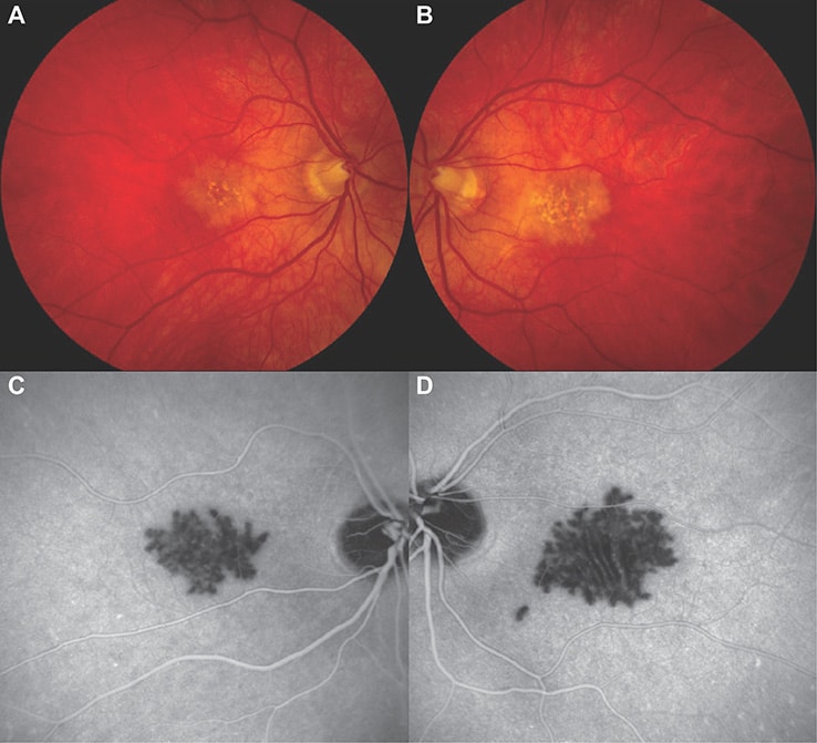
A 60-year-old male presented with headache, fatigue, and 6 weeks of blurry vision secondary to persistent placoid maculopathy. Colour fundus photographs demonstrated yellow-white placoid lesions in the macula OU with associated pigmentary changes (Fig 1A-B). Late-phase indocyanine-green angiography (ICG-A) revealed striking well-demarcated hypocyanescent regions corresponding to the macular lesions noted on colour fundus photography OU (Fig 1C-D).