Department of Ophthalmology, HD Hospital, Sherbrooke University, Sherbrooke, Quebec
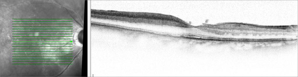
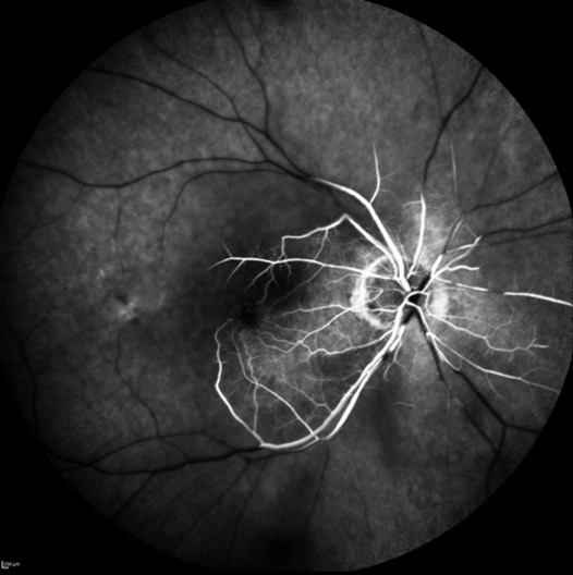
A 80-year-old female experimented nasal visual field loss in her right eye (OD) with preserved temporal visual field. Her past medical history was significant for age-related macular degeneration and hypertension. Best-corrected visual acuity was 6/6 OD and 6/9 OS. A pupillary afferent defect was present OD. Slip lamp examination was unremarkable in both eyes. Fundus examination OD revealed a whitened posterior pole with sparing in the papillomacular bundle. Spectral-domain optical coherence tomography OD revealed inner retinal hyperreflectivity with fovea sparing (Fig 1A). Fluorescein angiography showed a central retinal artery occlusion (CRAO) with cilioretinal sparing OD (Fig 1B).
Cilioretinal artery is an anatomic variant found in 15 to 30% of the population that often supplies the papillomacular bundle, but that supplies the foveola in only 10% of eyes1, 2. Therefore, its presence and specifically its distribution makes it possible of maintaining central vision in the event of a CRAO2, 3, 4.
1 Biousse V, Newman N. Retinal and optic nerve ischemia. Continuum (Minneap Minn). 2014 Aug. 20 (4 Neuro-ophthalmology):838-56.
2 Lorentzen SE. Incidence of cilioretinal arteries. Acta Ophthalmol (Copenh). 1970. 48 (3):518-24.
3 Pirasath S, Suganthan N, Malaravan M, Cilioretinal Artery Sparing Central Retinal Artery Occlusion, Austin Journal of Clinical Ophthalmology, 2015; 2(5): 1059
4 Hayreh SS, Zimmerman MB. Central retinal artery occlusion: visual outcome. Am J Ophthalmol. 2005;140:376-391.