Department of Ophthalmology, QEII Hospital, Dalhousie University, Halifax, Nova Scotia
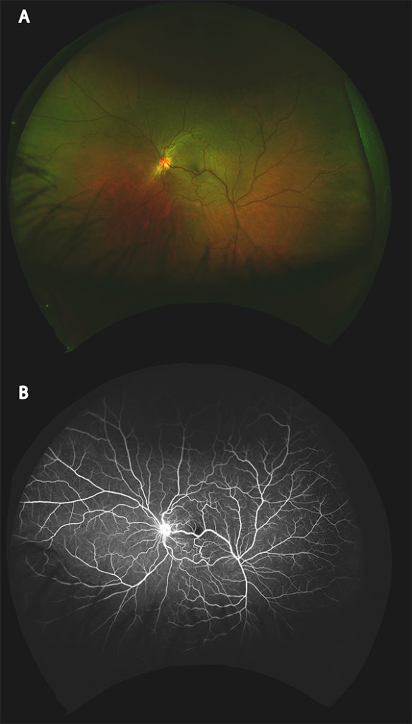
A 49-year-old male presented after blunt trauma OS. His past medical history was remarkable for blunt ocular trauma OD. Best-corrected visual acuity was 6/15 OD and 6/12 OS. Slit lamp examination of the anterior segment revealed traumatic mydriasis and an area of zonular insufficiency nasally from previous trauma OD. Slit lamp examination was unremarkable OS. Fundus examination OD revealed chronic chorioretinal scarring from previous trauma. Fundus examination OS revealed a retinal macrovessel extending through the macula and just inferior to the fovea. This finding was confirmed using fluorescein angiography (Figure 1A and B).
Congenital retinal macrovessels are large anomalous vessels that cross the horizontal raphe1,2. This is a rare finding3 that has been associated with decreased visual acuity and serous retinal detachments4.
1de Crecchio G, Mastursi B, Alfieri MC, Pignalosa B. Congenital retinal macrovessel. Ophthalmologica. 1986;193:143-5.
2Jager R, Timothy N, Joseph M, et al. Congenital Retinal Macrovessel. Retina. 2005;25(4):538-540.
3Hayasaka S, Katsube T, Ugomori S, Setogawa T. Abnormally Distributed Branches of the Retinal Vessels, Enlarged Macular Arteries and Long Cilioretinal Arteries. Ophthalmologica. 1990;200:194-197.
4Arai J, Kasuga Y, Koketsu M, Yoshimura N. Development and spontaneous reolution of serous retinal detachment in a patient with a congenital retinal macrovessel. Retina. 2000;130:527-528.